


What is Omphalocele?
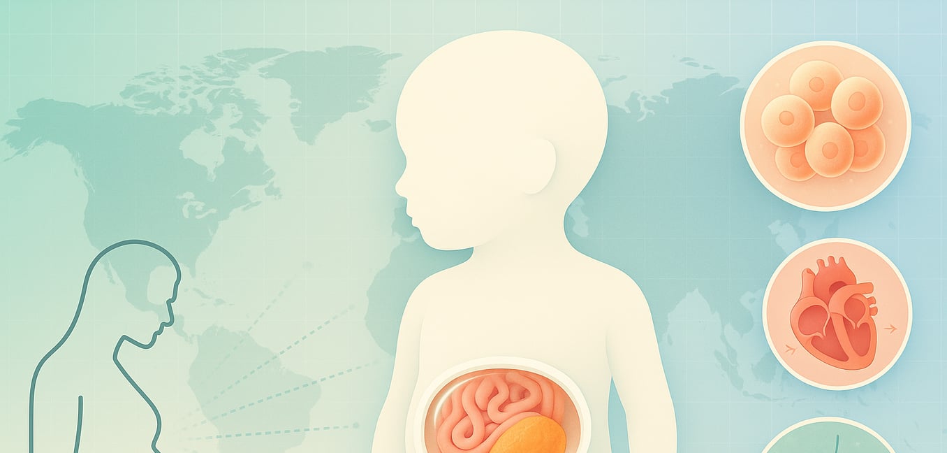
Omphalocele is a rare birth defect of the abdominal wall where an infant's internal organs—such as the intestines, liver, and stomach—protrude outside the body through an opening at the base of the umbilical cord. This condition originates between the 6th and 12th weeks of pregnancy when the intestines, which normally extend into the umbilical cord as they grow, fail to return to the abdominal cavity.
A key feature of an omphalocele is the presence of a protective sac that encloses the external organs. This sac, formed from a layer of the peritoneum (the membrane lining the abdominal cavity), shields the organs from direct exposure to amniotic fluid. This protective covering is a primary distinction from a similar condition, gastroschisis, where the intestines protrude without any sac.
Omphaloceles vary greatly in size, which significantly impacts treatment and prognosis. A small defect may contain only a single loop of intestine, while a "giant" omphalocele, defined as larger than 5 centimeters, can contain most of the abdominal organs. The size is critical because a larger defect often means the infant's abdominal cavity is underdeveloped and too small to accommodate the organs, complicating surgical repair.
Associated Conditions That Impact Prognosis
An omphalocele diagnosis is often a sign of more complex health issues. These associated anomalies can have a greater impact on an infant's long-term health than the abdominal wall defect itself. A thorough evaluation is essential because the presence of these conditions directly influences survival rates and treatment decisions.
Heart defects are the most common co-occurring issue, affecting up to 50% of infants with omphalocele. These can range from simple holes between the heart's chambers to more complex structural problems like Tetralogy of Fallot, a condition involving four related heart defects that disrupt normal blood flow. The risk for a cardiac anomaly increases with the size of the omphalocele, making a fetal echocardiogram (a detailed ultrasound of the heart) a standard part of the diagnostic process.
A significant number of infants with an omphalocele also have chromosomal abnormalities, such as Trisomy 18 (Edwards syndrome) or Trisomy 13 (Patau syndrome), both of which are associated with a very poor prognosis. Omphalocele can also be a key feature of specific genetic syndromes, most notably Beckwith-Wiedemann syndrome, which is characterized by an omphalocele, an unusually large tongue (macroglossia), and a larger-than-average body size (macrosomia).
Lung development can also be severely affected, especially in cases of a giant omphalocele. When a large portion of the liver is outside the abdomen, there is less space in the chest for the lungs to grow, leading to a condition called pulmonary hypoplasia (underdeveloped lungs). This can cause severe breathing problems and pulmonary hypertension (high blood pressure in the lungs) after birth.
Global Prevalence of Omphalocele and Why Rates Vary
Globally, omphalocele is a rare condition, with an estimated prevalence ranging from 1 to 4 cases for every 10,000 live births. However, this figure does not tell the whole story. The reported rate varies significantly across different populations and studies due to a combination of biological, medical, and societal factors.
The Discrepancy Between Prenatal and Live Birth Rates
One of the most striking statistical findings is the significant difference between the number of omphalocele cases detected during pregnancy and the number recorded among live-born infants. The incidence found on routine prenatal ultrasounds is much higher than the prevalence at birth. This gap is primarily explained by two factors directly linked to the high rate of associated anomalies.
First, the presence of severe chromosomal abnormalities or complex heart defects makes many affected pregnancies biologically non-viable. This leads to a higher rate of spontaneous miscarriage or stillbirth, meaning a substantial number of pregnancies diagnosed with omphalocele do not naturally progress to a live birth.
Second, in regions where it is legally and culturally permissible, some families may choose to terminate the pregnancy. This difficult decision is often made after counseling reveals a poor prognosis for the infant due to life-limiting chromosomal disorders or other severe defects. This practice directly reduces the number of live births with omphalocele, contributing to the lower prevalence rates seen at birth compared to prenatal screenings.
The Role of Maternal Age
Maternal age is one of the most consistently identified risk factors for omphalocele. The risk follows a U-shaped pattern, with an increased prevalence observed in pregnancies of both very young mothers (under 20 years old) and those of advanced maternal age (over 35). While the exact biological mechanisms are still under investigation, it is believed to be related to factors that can affect early cell division and embryonic development.
Geographical Differences and Data Reporting
The way omphalocele data is collected and reported creates notable variations in prevalence rates across different countries and regions.
The availability of prenatal screening and the legal status of pregnancy termination are major contributors to these differences. In countries with widespread access to advanced ultrasound and legal termination, the prevalence of omphalocele among live births is often lower because a higher proportion of severe cases do not reach term. In contrast, in nations with legal restrictions on termination for congenital abnormalities, a greater number of affected pregnancies are carried to term, resulting in a higher reported prevalence at birth.
The quality of national health registries also plays a crucial role. Regions with robust, population-based birth defect surveillance programs provide more accurate data than those with limited healthcare infrastructure, where underreporting is common. Furthermore, statistics from specialized tertiary care hospitals can show an artificially high prevalence. These centers receive high-risk pregnancy referrals from a wide geographical area, concentrating cases and creating a local "hotspot" that does not reflect the rate in the general population.
