

What is Noonan Syndrome?
Noonan syndrome is a genetic condition that affects how different parts of the body develop, leading to a wide range of distinct features and health issues7. It is caused by a mutation in one of several genes that regulate cell growth and division7. This change can be passed down from a parent or occur for the first time in an individual7.
Because the effects vary greatly from person to person, the condition can be mild or more severe37. Some of the most common characteristics include:
- Distinctive facial features: This includes wide-set, downward-slanting eyes with droopy lids, low-set ears, and a short or "webbed" neck7. These traits often lead to a diagnosis and may become less noticeable with age.
- Heart conditions: Because heart conditions are common, doctors pay close attention to the baby’s heart during pregnancy and after birth. The most frequently seen issues are pulmonary valve stenosis (a narrowing of a heart valve) and hypertrophic cardiomyopathy (a thickening of the heart muscle), both of which can be monitored and managed by specialists7.
- Growth and development: While birth weight is often typical, growth may slow over time, leading to short stature in adulthood72. Children may also experience developmental delays, though most have normal intelligence57.
- Other physical traits: Many people may bruise or bleed easily7. Other possible signs include an unusually shaped chest, undescended testicles in males, and issues with the lymphatic system that cause swelling73.
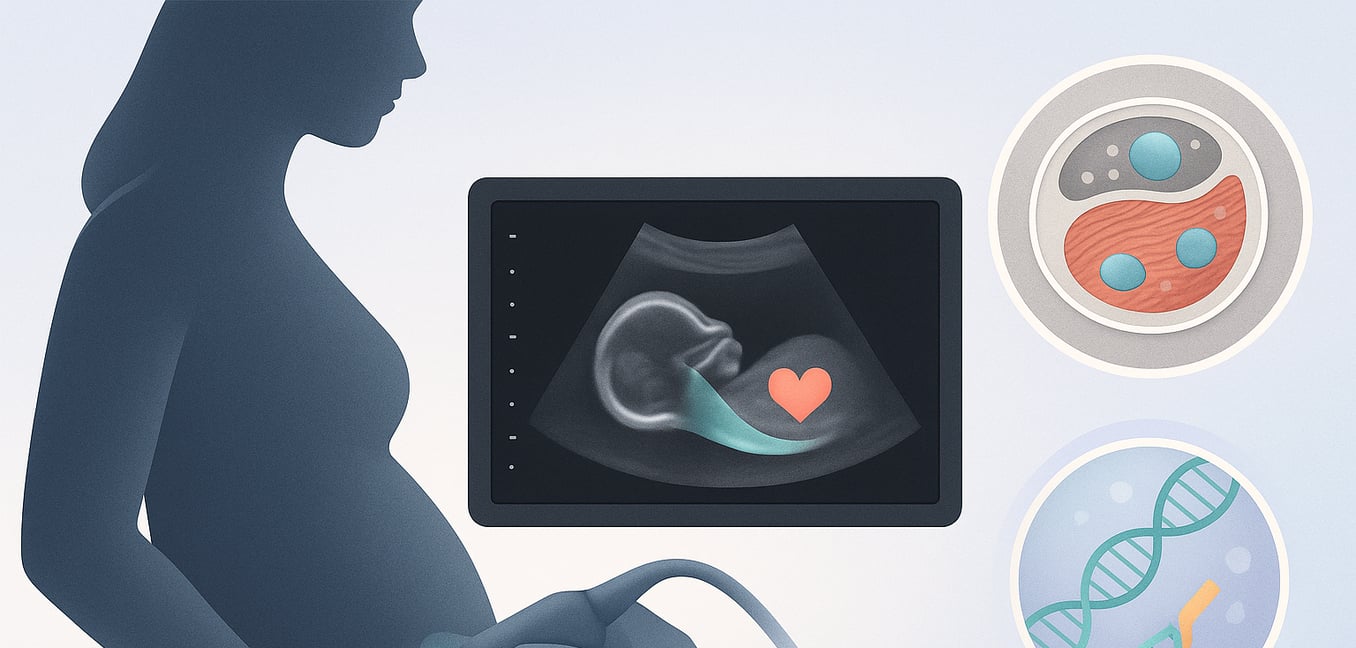
How Ultrasound Can Suggest Noonan Syndrome
While Noonan syndrome can only be diagnosed with genetic testing, clues found during routine prenatal ultrasound scans can be the first sign that suggests the condition5. An ultrasound cannot provide a definite answer, but it can reveal a pattern of physical markers that may prompt a recommendation for further testing5.
Fluid-Related Markers
The most common signs seen on an ultrasound are related to the body's lymphatic system, which is responsible for draining fluid3.
- In the first trimester, this may appear as an increased nuchal translucency (NT), which is a measurement of the fluid-filled space at the back of the baby’s neck.
- Later in pregnancy, lymphatic issues can lead to a cystic hygroma (a fluid-filled sac on the neck) or a buildup of fluid around the lungs, heart, or in the abdomen57.
- In severe cases, this can cause widespread swelling throughout the baby’s body, a condition known as hydrops fetalis5.
This spectrum of fluid-related findings is a hallmark prenatal feature of Noonan syndrome5.
Heart Conditions
Specific observations about the baby’s heart are also significant3. The two most characteristic heart conditions are pulmonary stenosis and hypertrophic cardiomyopathy. A major challenge is that these issues can be subtle or may only develop late in pregnancy4. A fetal heart scan at 20 weeks might appear normal, but the heart muscle could thicken or a valve could narrow in the third trimester2. For this reason, if Noonan syndrome is suspected due to other markers, a specialized fetal heart ultrasound (echocardiogram) and follow-up scans are often recommended5.
Other Potential Clues
A cluster of other findings can strengthen the suspicion of Noonan syndrome, especially when seen alongside fluid or heart concerns5.
- Polyhydramnios: An excess of amniotic fluid is a common finding, particularly in the second half of pregnancy83.
- Other markers: Less frequent but still important signs include mild swelling in the baby's kidneys (pyelectasis) or the presence of only one artery in the umbilical cord instead of the usual two4.
While any of these signs alone may not be a major concern, their presence together creates a compelling pattern that points toward the need for further investigation3.
Confirming the Diagnosis with Genetic Testing
When ultrasound scans reveal features suggestive of Noonan syndrome, genetic testing is the only way to confirm the diagnosis before birth5. This process analyzes the baby’s DNA for the specific gene mutations that cause the condition, providing parents and their medical team with a clear answer to help them prepare for the baby’s needs37.
The diagnostic journey typically involves these key steps:
- Targeted Gene Testing: A specialized test looks for mutations in the group of genes known to cause Noonan syndrome and related conditions85. This is necessary because the syndrome can be caused by changes in many different genes, though the PTPN11 gene is the most common8.
- Fetal Sample Collection: To analyze the baby's DNA, a sample is gathered through either chorionic villus sampling (CVS), usually done between 10 and 13 weeks, or amniocentesis, performed after 15 weeks85. A doctor will discuss the small risks associated with these procedures57.
- Interpreting the Results: If the test finds a specific gene change known to cause Noonan syndrome, the diagnosis is confirmed83. A negative result makes the diagnosis much less likely but doesn’t completely rule it out, as the mutation could be in a gene not yet identified or included on the test panel3.
- Genetic Counseling: Experts help parents understand the testing process, the meaning of the results, and what to expect from a diagnosis73. This guidance is crucial for making informed decisions about pregnancy management and future care.
Challenges and Limitations in Prenatal Detection
Despite advances in medical imaging, identifying Noonan syndrome before birth remains challenging4. The condition’s wide variability is the primary reason a diagnosis can be missed, even by experienced specialists83.
Navigating the path to a prenatal diagnosis comes with several inherent difficulties:
- Subtle or Absent Signs: Many fetuses with Noonan syndrome show no clear markers on an ultrasound, especially in milder cases48. A baby might have normal growth and no obvious fluid or heart issues, meaning the diagnosis is often missed before birth4.
- Non-Specific Markers: Signs like increased nuchal translucency or excess amniotic fluid can be linked to many other conditions, such as Down syndrome or maternal diabetes3. This overlap makes it difficult to pinpoint Noonan syndrome without genetic testing8.
- Late-Appearing Features: Some of the most characteristic signs may only develop late in pregnancy5. For example, hypertrophic cardiomyopathy often becomes visible in the third trimester, long after the standard mid-pregnancy anatomy scan is complete4.
