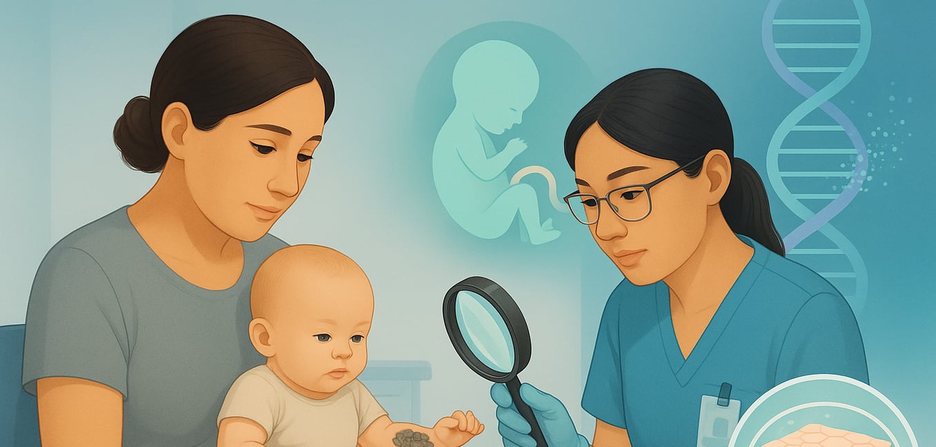


What is Recessive X-Linked Ichthyosis?
Recessive X-linked ichthyosis (XLI) is an inherited skin disorder that almost exclusively affects males, typically becoming apparent shortly after birth. It is a lifelong condition characterized by dry, scaly skin, but its impact can extend beyond the surface.
The Genetic Cause and Inheritance
XLI stems from a deficiency of an enzyme called steroid sulfatase (STS). In over 90% of cases, this deficiency is caused by the complete deletion of the STS gene, which resides on the X chromosome.
Because males have only one X chromosome (XY), inheriting a single altered copy from their mother is sufficient to cause the condition. Females (XX), on the other hand, have a second, functional X chromosome that compensates for the faulty one. As a result, they are typically unaffected carriers who can pass the gene deletion to their sons.
Key Physical and Associated Features
The most visible sign of XLI is persistent, widespread skin scaling. The scales are often dark brown or grey, polygonal in shape, and most prominent on the neck, torso, and the outer surfaces of the arms and legs. The palms and soles are usually spared.
The absence of the STS enzyme disrupts the normal shedding of skin cells, causing dead cells to build up and form thick, adherent scales. Beyond the skin, this enzyme deficiency is linked to other health issues. A significant number of individuals with XLI have an increased risk of cryptorchidism, where one or both testicles fail to descend. Many also develop harmless, dot-like opacities in the cornea of the eye, which do not typically affect vision. The placental enzyme deficiency can also lead to a delay in the onset of labor for mothers carrying an affected male fetus.
Initial Clues from Prenatal Screening
The first indication of XLI during a pregnancy often comes from a routine blood test offered to expectant mothers. This maternal serum screening, while not designed to detect XLI specifically, can reveal hormonal irregularities that act as a powerful signpost for the condition in a male fetus.
The key finding is a significantly low, or even undetectable, level of a hormone called unconjugated estriol (uE3). The placenta of a male fetus with XLI lacks the STS enzyme, which is essential for producing estrogens like estriol. This blockage in the hormonal pathway results in a profound uE3 deficiency in the mother’s blood.
While maternal serum screens (often called the "triple" or "quad" screen) primarily assess risk for conditions like Down syndrome, an extremely low uE3 level, especially when other markers are normal, is a classic hallmark of placental STS deficiency. This finding strongly prompts healthcare providers to consider XLI and recommend more definitive diagnostic testing.
Definitive Genetic Diagnosis and Its Implications
While low estriol is a strong hint, a definitive diagnosis requires analyzing the fetus’s genetic makeup. Advanced prenatal tests can precisely identify the underlying changes in the STS gene, providing families with a clear answer and a detailed understanding of the diagnosis.
Primary Diagnostic Tools
The gold standard for confirming XLI is a chromosomal microarray (CMA). This high-resolution test examines the fetus’s chromosomes for small missing or extra pieces of DNA. Since most XLI cases are caused by a complete deletion of the STS gene, CMA is exceptionally effective. A major advantage of CMA is its ability to determine the exact size of the deletion, revealing whether adjacent genes are also missing. This is crucial for identifying the risk of a more complex contiguous gene syndrome, which can involve additional health issues.
Another reliable method is fluorescence in situ hybridization (FISH). This technique uses fluorescent probes that are designed to bind specifically to the STS gene. In a sample of fetal cells, the absence of a fluorescent signal at the expected location on the X chromosome provides clear, visual proof that the gene is deleted. FISH is a focused and relatively rapid way to confirm the most common cause of XLI.
Investigating Rarer Cases
In the small fraction of cases where XLI is suspected but CMA and FISH results are normal, the cause is likely a point mutation—a tiny error within the STS gene’s DNA sequence. These subtle changes are too small for microarray or FISH to detect. In this scenario, STS gene sequencing is performed. This analysis reads the gene’s code letter by letter to pinpoint the specific error preventing the enzyme from functioning correctly.
The Value of a Precise Diagnosis
Genetic testing provides more than a simple yes-or-no answer. It is the definitive method for confirming carrier status in the mother and other female relatives. While a mother of an affected son is an obligate carrier, genetic confirmation is vital for her future pregnancies and for counseling her sisters or daughters. This knowledge empowers family members to make informed reproductive choices.
Distinguishing XLI from Other Types of Ichthyosis
Differentiating XLI from other inherited scaling disorders is a critical diagnostic step, as the inheritance pattern, associated health risks, and management strategies vary significantly between conditions.
Ichthyosis Vulgaris (IV)
Ichthyosis vulgaris is the most common form of ichthyosis but is typically milder than XLI. The scales in IV are usually fine and white, in contrast to the larger, darker, polygonal scales of XLI. A key difference is that IV characteristically spares the flexural areas (like the creases of the elbows and knees), which are often affected in XLI. Most importantly, IV is an autosomal dominant condition, meaning it can be passed from an affected parent to children of either sex.
Lamellar Ichthyosis
A form of autosomal recessive congenital ichthyosis (ARCI), lamellar ichthyosis presents a more severe picture from birth. Newborns are often encased in a tight, transparent film known as a collodion membrane, which is not a feature of XLI. After the membrane sheds, it reveals large, dark, plate-like scales across the entire body. As an autosomal recessive disorder, it occurs when a child inherits a faulty gene from both unaffected carrier parents, pointing genetic testing toward genes like TGM1 rather than STS .
Harlequin Ichthyosis (HI)
Harlequin ichthyosis is the rarest and most severe form of ARCI. Its presentation at birth is dramatically different from XLI. Infants with HI are born with thick, armor-like plates of skin separated by deep fissures, a condition that is often life-threatening without immediate, intensive medical care. It is an autosomal recessive condition caused by mutations in the ABCA12 gene. Differentiating it from XLI is essential, as the prognosis and level of required neonatal care are vastly different.
