


The Cornea's Clear Foundation: The Role of Collagen
The human cornea is a biological marvel, acting as both a tough, protective shield for the inner eye and a perfectly clear window for light to pass through. This unique combination of strength and transparency is primarily due to its main structural protein, collagen. The cornea's middle layer, the stroma, makes up about 90% of its total thickness and is composed almost entirely of this vital protein. The health of this collagen framework is directly linked to the cornea's ability to function, and when it fails, debilitating conditions like corneal dystrophies can arise.
The Architecture of a Healthy Cornea
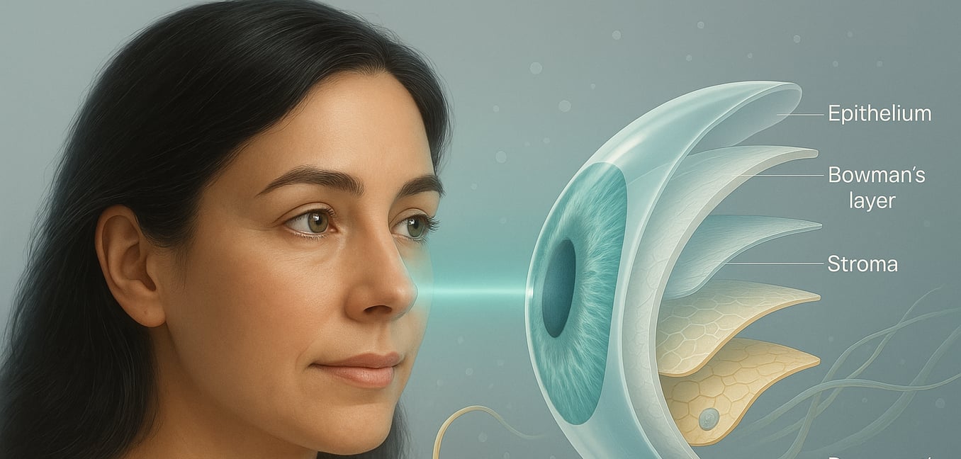
To understand what happens in disease, we must first appreciate the intricate design of a healthy cornea. Its structure is a masterpiece of biological engineering, with each layer performing a specific role supported by a collagen-based framework.
The Central Collagen Powerhouse
The stroma is the cornea's main structural component. It consists of hundreds of incredibly thin layers, or lamellae, which are stacked on top of each other with remarkable precision, much like sheets of plywood. Each lamella is packed with precisely aligned type I collagen fibrils. The fibrils in each layer run at an angle to those in the layers above and below, creating a cross-hatched, lattice-like arrangement. This design is the secret to the cornea's immense strength, while the uniform spacing of the fibrils minimizes light scatter, ensuring vision remains crystal clear.
The Protective Barrier of Bowman's Layer
Just beneath the cornea's surface epithelium lies Bowman's layer, a dense and tough sheet composed of randomly arranged collagen fibrils. Unlike the highly organized stroma, this layer contains no cells and cannot regenerate if significantly damaged. Its main purpose is to serve as a strong protective barrier, shielding the underlying stroma from injury. It also provides a smooth, stable foundation upon which the surface epithelial cells can anchor and grow, acting as a critical first line of defense for the deeper corneal structures.
The Endothelium's Hexagonal Foundation
The deepest collagen-based layer is the Descemet membrane, which serves as a specialized basement membrane for the vital endothelial cells. In a healthy cornea, this membrane is not just a simple sheet. It possesses a delicate and consistent pattern of fine hexagonal structures, creating a unique honeycomb-like scaffold. This architecture, built from a protein called Type VIII collagen, provides the perfect combination of flexibility and support for the endothelial cells, allowing them to effectively perform their crucial pump function.
Corneal Dystrophy: When the Foundation Crumbles
When this delicate structural integrity is compromised by genetic factors, a group of conditions known as corneal dystrophies can develop. One of the most common is Fuchs' endothelial dystrophy, a slowly progressing disease that provides a clear example of how disruptions in the cornea's deepest layers can lead to significant vision loss.
The Origin of Disruption
The hallmark of Fuchs' dystrophy is the gradual appearance of tiny, wart-like growths called guttata on the Descemet membrane. These growths are not foreign invaders but are abnormal deposits of fibrillar collagen and other materials produced by the distressed endothelial cells themselves. As these mushroom-shaped deposits accumulate, they create a bumpy, uneven surface often described as having a "beaten metal" appearance. This fundamentally alters the structural foundation upon which the crucial endothelial cell layer rests.
A Failing Pump System
The corneal endothelium works like a microscopic pump, constantly removing fluid from the stroma to keep the cornea thin and transparent. The progressive formation of guttata stretches and eventually destroys these hard-working endothelial cells, leading to a steady decline in their numbers. While the remaining cells enlarge to cover the gaps, this patchwork solution cannot maintain the powerful pump function. As the system fails, the cornea becomes waterlogged, or edematous. This swelling separates the neatly arranged collagen fibrils in the stroma, causing the cloudiness and blurred vision characteristic of the disease.
A Closer Look: How Collagen Failure Drives Fuchs' Dystrophy
In Fuchs' dystrophy, the elegant hexagonal collagen scaffold of the Descemet membrane undergoes a destructive transformation. This is an active, pathological remodeling process where the cornea creates an entirely new and dysfunctional foundation, which drives the disease forward.
First, the fine, physiological hexagonal structure that provides a flexible anchor for endothelial cells begins to break down. This delicate network loses its consistency, showing areas of thinning and discontinuity. This initial breakdown compromises the stable platform for the endothelial cells, contributing to their stress and eventual death.
In response to this damage, the cornea initiates a flawed repair process. It begins depositing thick bundles of fibrillar collagen where they do not belong. This abnormal collagen organizes into a dense, honeycomb-like network that entwines around the guttata, creating a thick and disorganized layer. This pathological network is not a restoration of the original structure but a scar-like reaction that further worsens the condition.
This newly formed fibrous wall creates significant functional problems. Its density can contribute to the cornea's cloudiness and worsen the fluid buildup, leading to greater corneal thickness. This scar-like barrier is also thought to prevent healthy endothelial cells from the periphery from migrating toward the damaged central area, hindering any natural repair.
The Genetic Blueprint for Disease
The development of Fuchs' dystrophy is often not a matter of chance; the blueprint for the condition can be found within an individual's genetic code. For many, specific mutations in genes responsible for corneal structure are the primary drivers, disrupting the normal processes of cellular maintenance and collagen production.
Direct Flaws in the Building Blocks
The COL8A2 gene provides a direct link to a form of Fuchs’ dystrophy that appears earlier in life. This gene holds the specific instructions for producing a component of type VIII collagen, the very protein that forms the delicate hexagonal foundation of the healthy Descemet membrane. When this genetic blueprint contains an error, the resulting collagen is defective. This compromises the membrane's architecture from the start, a classic example of how a flaw in a structural component can initiate disease.
Errors in Site Management
The most common genetic factor for late-onset Fuchs' dystrophy is found in the TCF4 gene. Unlike COL8A2, which provides the blueprint for a single part, TCF4 acts like a construction site manager. It is a "transcription factor," meaning it directs the activity of many other genes involved in building and maintaining the cornea. The most common mutation is not a simple typo but a "stutter" in the genetic code—a repetition of a three-letter DNA sequence—which disrupts the manager's instructions and significantly increases the lifetime risk of developing the disease.
A Complex Genetic Picture
The story is further complicated by other genes, such as SLC4A11 and ZEB1, which are responsible for different cellular jobs, from transporting ions to guiding cell specialization. This genetic complexity helps explain why many cases do not follow a clear inheritance pattern and may arise from a combination of multiple subtle genetic risks. It underscores that the cornea’s clarity depends on a wide range of perfectly functioning and interconnected systems.
