


Understanding Corneal Dystrophy
Corneal dystrophies are a family of genetic disorders that affect the cornea, the clear front window of the eye. These conditions are caused by mutations in specific genes that disrupt the normal function and health of corneal cells. This genetic flaw leads to the gradual buildup of abnormal material within one or more of the cornea's distinct layers.
Over time, this accumulation compromises the cornea's transparency, scattering light and causing a range of vision problems. The specific symptoms depend on which layer is affected. If the outer layer (epithelium) has deposits, it can cause painful recurrent corneal erosions. In contrast, issues in the deeper layers typically lead to painless but progressive clouding of vision. Because the conditions are hereditary, understanding a patient's family history is often a key part of diagnosis.
Managing Symptoms with Non-Surgical Treatments
Before considering surgery, various non-surgical strategies can effectively manage the daily symptoms of corneal dystrophy. These treatments focus on providing comfort, protecting the corneal surface, and maintaining the best possible vision.
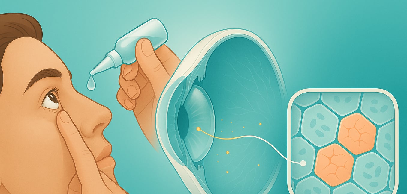
Specialized Eye Drops and Ointments
To manage daily symptoms, your doctor may prescribe targeted eye drops. For the gritty, foreign-body sensation common in surface-level dystrophies, preservative-free lubricating drops can provide relief. For conditions that cause corneal swelling, such as Fuchs’ dystrophy, hypertonic saline (5% sodium chloride) drops and ointments are used. These solutions draw excess fluid from the cornea, helping to reduce the foggy vision that is often worse in the morning.
Therapeutic Contact Lenses
In cases where recurrent corneal erosions cause significant pain, a bandage contact lens can act as a protective shield. This special soft lens covers the cornea, preventing the eyelid from rubbing against the delicate surface with every blink. This not only reduces pain but also creates a stable environment that promotes faster healing. Antibiotic eye drops are often prescribed alongside the lens to prevent infection.
Corrective Eyewear
While glasses and standard contact lenses cannot stop the progression of the dystrophy, they are essential for maximizing vision. The deposits or swelling in the cornea can distort its surface, creating refractive errors like astigmatism. A proper prescription for glasses can help refocus light more accurately onto the retina. For some, rigid gas permeable (RGP) contact lenses may offer superior clarity by creating a new, perfectly smooth front surface for the eye.
Consistent Monitoring and Proactive Care
Since most corneal dystrophies progress slowly, regular follow-up appointments with your eye specialist are crucial. These visits allow your doctor to monitor changes in your vision and corneal health, ensuring your treatment plan is optimized. For children, this monitoring is especially important to prevent the development of amblyopia ("lazy eye"), which can cause permanent vision loss if not addressed early.
Surgical Interventions: Restoring Corneal Clarity
When non-surgical treatments no longer provide clear vision or adequate comfort, surgery becomes the most effective option. Modern techniques can restore corneal clarity by resurfacing the cornea with a laser or replacing only the damaged tissue with a transplant.
Excimer Laser Phototherapeutic Keratectomy (PTK)
For dystrophies affecting the cornea's superficial layers, like Map-Dot-Fingerprint or Lattice dystrophy, PTK is an excellent, minimally invasive option. This procedure uses a cool ultraviolet laser to precisely remove microscopic deposits and smooth out surface irregularities. By clearing the material that causes cloudiness and painful erosions, PTK can significantly improve vision and comfort, often delaying or preventing the need for a transplant.
Partial-Thickness Corneal Transplant (Lamellar Keratoplasty)
This advanced approach allows surgeons to replace only the diseased layers of the cornea while leaving healthy tissue intact. For Fuchs’ dystrophy, which affects the innermost endothelial layer, a procedure called Descemet Membrane Endothelial Keratoplasty (DMEK) is common. The surgeon removes the faulty endothelial layer and replaces it with a healthy donor layer. Because most of the patient's own cornea remains, this technique offers faster visual recovery and a lower risk of tissue rejection.
Full-Thickness Corneal Transplant (Penetrating Keratoplasty)
This traditional transplant is recommended when a dystrophy has caused scarring across all layers of the cornea. During this procedure, the surgeon removes the entire central portion of the damaged cornea and replaces it with a matched donor cornea, which is secured with ultra-fine stitches. While highly successful at restoring sight, this surgery involves a longer recovery period and a higher risk of graft rejection, which is managed with long-term steroid eye drops.
Future and Emerging Therapies
Ophthalmology is rapidly advancing toward a future that may not rely on donor tissue. Researchers are exploring groundbreaking technologies that aim to regenerate corneal tissue or encourage the eye to heal itself, promising treatments that are less invasive and more accessible.
Bioengineered Corneas
To address the worldwide shortage of donor corneas, scientists are developing "bioengineered" corneas in labs. These are created using biocompatible polymers or purified collagen. Some teams are even using advanced bioprinting technology to construct new tissue layer by layer, complete with living cells. The goal is to create a readily available, off-the-shelf solution that can eliminate transplant waiting lists.
Cell-Based Regeneration
Instead of transplanting tissue, this revolutionary approach uses healthy, lab-grown cells injected directly into the eye. This is particularly promising for Fuchs’ dystrophy, where endothelial cells fail. Researchers can now cultivate these specialized cells in a lab, allowing cells from a single donor to be multiplied to treat many patients. Once injected, these cells get back to work pumping fluid from the cornea, restoring clarity with a potentially lower risk of immune rejection.
Promoting Natural Repair
An elegant new technique, known as Descemet Stripping Only (DSO), helps the eye heal itself. A surgeon gently removes only the diseased central endothelial cells, leaving the healthy peripheral cells intact. The patient then uses special eye drops containing a "ROCK inhibitor," a compound that signals the remaining healthy cells to migrate and repopulate the cleared area. This strategy avoids the need for donor tissue entirely, eliminating the risk of rejection.
