


What is Omphalocele?
Omphalocele, sometimes called exomphalos, is a congenital birth defect of the abdominal wall where some of a baby's abdominal organs protrude through an opening at the base of the umbilical cord. This occurs very early in fetal development when the intestines, which normally extend into the umbilical cord and then return to the abdomen, fail to complete this final step.
A defining characteristic of an omphalocele is that the protruding organs are contained within a thin, translucent sac. This protective covering is a key feature that helps doctors distinguish it from a similar condition, gastroschisis. In gastroschisis, there is no protective sac, and the intestines protrude through a hole next to the umbilical cord, leaving them exposed directly to the amniotic fluid.
The size of an omphalocele can vary significantly, from a small defect containing only a loop of the intestine to a "giant" omphalocele larger than five centimeters that includes the liver. Because omphalocele is frequently associated with other genetic and chromosomal conditions, a diagnosis prompts a comprehensive evaluation to create a complete picture of the baby's health.
Prenatal Diagnosis
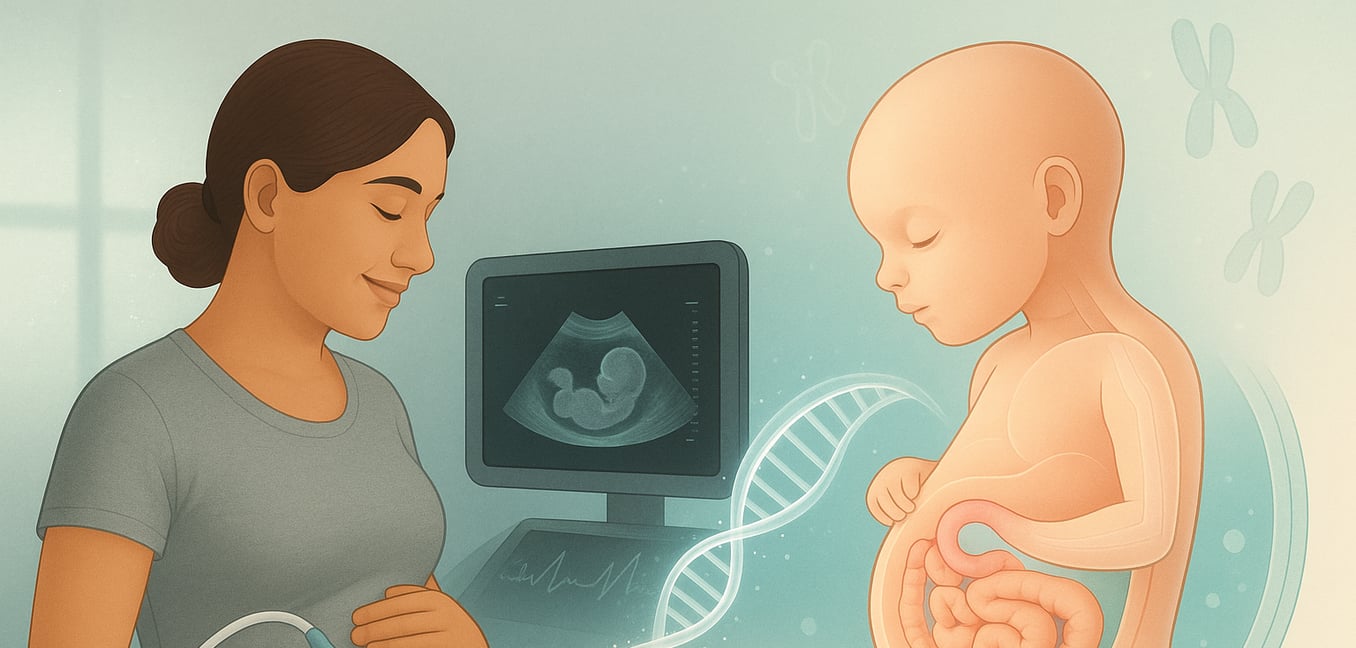
An omphalocele is most often diagnosed during pregnancy, typically with a routine ultrasound around the 20-week mark. This early detection allows the medical team and family to prepare a detailed plan for delivery and postnatal care. The prenatal evaluation involves several key steps to understand the baby's specific condition.
Detailed Ultrasound
A high-resolution, Level II ultrasound provides a detailed look at the baby’s anatomy. This scan is used to:
- Confirm the omphalocele: By identifying the protective sac at the base of the umbilical cord, its size, and which organs (like the liver or intestines) are inside.
- Screen for other issues: By performing a full-body scan to check for other common structural differences, especially in the heart. A specialized fetal echocardiogram is often performed to get a closer look at the heart's structure and function.
- Assess lung development: To check for pulmonary hypoplasia (underdeveloped lungs), a common concern in babies with large omphaloceles. The organs outside the abdomen can restrict chest growth, leaving less space for the lungs to develop.
Advanced Imaging and Genetic Testing
To gather more detailed information, the care team may recommend further testing:
- Fetal MRI: This imaging technique provides a highly detailed view of the baby’s body without using radiation. It is particularly useful for measuring the baby’s lung volume more precisely. By measuring the lungs, doctors can compare their size to what is expected for the baby's gestational age. This helps predict whether the baby will need breathing support after birth.
- Amniocentesis: This procedure involves taking a small sample of amniotic fluid to analyze the baby’s chromosomes. It can definitively identify or rule out associated genetic conditions, which is critical for understanding the baby’s overall prognosis and guiding care decisions.
- Genetic Counseling: A genetic counselor helps families understand the results of genetic tests. They explain what a diagnosis might mean for the baby’s health, discuss the chances of it happening in future pregnancies, and provide support to help families make informed decisions.
Postnatal Diagnosis and Initial Assessment
Immediately after birth, the medical team performs a thorough physical examination to confirm the diagnosis and assess the baby’s overall health. This initial evaluation guides the first steps of care.
The assessment focuses on three key areas:
- Confirming the Omphalocele: The team visually inspects the sac-covered mass at the umbilical cord, noting its size and whether the sac is intact or ruptured. This information is essential for planning the timing and type of surgical repair needed.
- Assessing Breathing and Heart Function: A top priority is checking the baby’s respiratory and cardiovascular stability. The team monitors breathing rate and oxygen levels to see if the baby needs support from a ventilator, especially if underdeveloped lungs (pulmonary hypoplasia) were a concern on prenatal scans.
- Checking for Signs of Associated Syndromes: The examination includes a head-to-toe check for physical markers that may suggest an underlying genetic condition. This includes looking for features like an enlarged tongue or specific ear creases, which can point toward a specific syndrome.
Understanding Associated Conditions
A crucial part of the diagnostic process is identifying any related syndromes, as they significantly impact the baby's overall care plan and long-term outlook. Omphalocele is strongly linked to several genetic and chromosomal conditions.
Beckwith-Wiedemann Syndrome (BWS)
BWS is one of the most common genetic syndromes associated with omphalocele. It is an overgrowth syndrome, and babies with BWS are often larger than average at birth (macrosomia). Other common features include an enlarged tongue (macroglossia), ear creases, and low blood sugar (hypoglycemia) in the newborn period. While children with BWS typically have normal cognitive development, they have an increased risk of developing certain childhood cancers. Because of this, they require a regular screening schedule with abdominal ultrasounds and blood tests for the first several years of life.
Chromosomal Conditions (Trisomies)
Omphalocele is also strongly associated with chromosomal abnormalities, most notably Trisomy 13 (Patau syndrome), Trisomy 18 (Edwards syndrome), and Trisomy 21 (Down syndrome). The presence of a trisomy profoundly affects a baby’s prognosis, as these conditions often involve severe heart defects and significant developmental challenges. This is why genetic testing via amniocentesis is so often recommended when an omphalocele is detected, as confirming or ruling out a chromosomal condition is fundamental to understanding the baby's future health needs and guiding family counseling.
