


New and Emerging Treatments for Fuchs' Dystrophy
Fuchs' Endothelial Dystrophy is a hereditary eye condition that affects the cornea, the clear front surface of the eye. It targets the endothelium, the cornea's innermost layer of "pump" cells responsible for maintaining its clarity. In Fuchs' Dystrophy, these pump cells begin to fail and disappear. As they do, the cornea can no longer pump out excess fluid, causing it to swell and become cloudy, like a window fogging up.
Early symptoms often include hazy vision, especially in the morning, which can be mistaken for cataracts. As the disease progresses, this fogginess can persist all day, leading to significant vision loss. For decades, the primary treatment has been a corneal transplant. Today, the gold standard is a partial-thickness procedure called Descemet’s Membrane Endothelial Keratoplasty (DMEK), where only the diseased inner layer is replaced with healthy donor tissue. While highly effective, new innovations are offering patients less invasive options and even the possibility of avoiding transplants altogether.
Innovations in Corneal Surgery
Even as DMEK remains a top treatment, surgeons are refining both transplant and transplant-free procedures to be safer, more effective, and accessible to more patients.
Descemet's Stripping Only (DSO): A Transplant-Free Approach
An innovative procedure known as Descemet's Stripping Only (DSO) harnesses the cornea's own remarkable ability to heal. In this delicate operation, a surgeon removes a small, four-millimeter circle of the diseased cell layer from the very center of the cornea, leaving the rest of the cornea untouched. No donor tissue is inserted. This "stripping" action creates a clean slate, encouraging the patient’s own healthy endothelial cells from the outer edges of the cornea to migrate inwards and repopulate the cleared area.
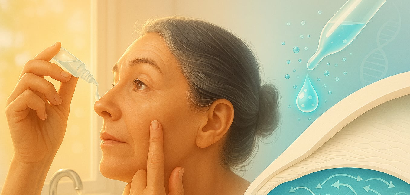
To boost this natural healing process, the procedure is now almost always supplemented with rho-kinase (ROCK) inhibitor eye drops. These drops act as a powerful catalyst, giving the healthy peripheral cells permission to move and multiply, dramatically speeding up corneal clearing and pushing success rates to over 90% for carefully selected patients.
The ideal candidate for DSO has a disease that is mostly confined to the central cornea, with a healthy reserve of cells in the periphery. Patients must also understand that vision is initially worse after surgery and typically takes one to three months to clear. If the cornea fails to clear on its own, a standard DMEK transplant is performed. The key benefit is the complete elimination of transplant rejection risk, which means patients can avoid the lifelong steroid eye drops required after a traditional transplant.
Advancing the Gold Standard: Better DMEK Techniques
The gold-standard DMEK procedure is also evolving. A key challenge in this surgery is the extreme fragility of the donor tissue, an endothelial layer as thin as plastic wrap. Protecting these delicate cells during preparation and implantation is critical for success.
To address this, new surgical tools have been developed to make the process safer. One such device, the DMEK EndoGlide, acts like a specialized cartridge, allowing the surgeon to gently and precisely guide the tissue into place, almost like sliding a delicate film into a protective sleeve. Studies show that this gives the surgeon more control, leading to a faster and more predictable implantation of the graft. For patients, a smoother, more controlled surgery reduces the risk of cell loss, which in turn improves the odds of a successful transplant and excellent long-term vision.
The Rise of Medical Therapy
Beyond surgery, a new class of medication is transforming how Fuchs' Dystrophy can be managed, offering hope for slowing the disease and improving outcomes for existing procedures.
ROCK Inhibitors as a Standalone Treatment
Originally developed for glaucoma, ROCK inhibitor eye drops (like netarsudil) have shown a remarkable ability to rejuvenate the cornea's endothelial cells. This has opened the door to using them as a standalone medical therapy. Clinical studies have shown that these drops can reduce corneal swelling and improve vision in patients with Fuchs' Dystrophy even without any surgery.
The goal of this approach is to directly improve the function of the patient's existing endothelial cells, potentially slowing or even halting the disease's progression. This could one day offer a non-invasive way for patients to manage their condition, delaying or possibly avoiding the need for surgery for many years. Major clinical trials are currently underway to compare the effectiveness of DSO with ROCK inhibitors against the gold-standard DMEK transplant.
On the Horizon: Regenerative Medicine and Gene Therapy
Looking ahead, the most exciting frontier in treating Fuchs' Dystrophy involves regenerative medicine that could one day make surgery obsolete. The goal is to restore a healthy endothelial layer with a simple injection of new cells, a concept moving rapidly from theory to clinical reality.
Injectable Cell Therapy
One leading approach involves cultivating endothelial cells in a laboratory. In this process, healthy cells are harvested from a single donor cornea and multiplied to create enough doses to treat as many as 100 patients. These lab-grown cells are then delivered via a simple injection into the front of the eye. After the injection, the patient lies face down for a few hours, allowing the new cells to settle and attach to the back of the cornea. This technology, already approved in Japan and in late-stage trials in the U.S., could solve the global shortage of donor corneas.
Magnetic Cell Delivery
An even more streamlined method uses magnetism to ensure the new cells get exactly where they need to go. In this technique, lab-grown endothelial cells are infused with tiny magnetic nanoparticles. After being injected into the eye, the patient wears a special magnetic patch externally for a short time. This magnetic field guides the cells to settle onto the cornea's inner surface without requiring the patient to lie face-down. The procedure is so simple it could potentially be done in a doctor's office, promising a quick recovery and wider accessibility.
Targeting the Source with Gene Therapy
Further in the future, researchers are targeting the genetic source of the disease itself. For most patients, Fuchs' is caused by a known flaw in the TCF4 gene. Gene therapy aims to intercept this flaw by injecting molecules designed to bind to the faulty genetic message and neutralize it before it can cause harm to the cells. While still in the very early stages of development, this strategy represents a paradigm shift from replacing damaged cells to preventing them from dying in the first place, offering the potential for a true cure.
