


What Is Corneal Dystrophy?
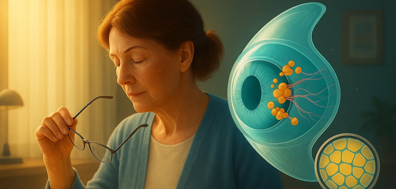
The cornea is the clear, dome-shaped window at the front of your eye, responsible for focusing light to create sharp images. Corneal dystrophies are a group of more than 20 genetic disorders that cause abnormal materials to build up in one or more of the cornea's five distinct layers. This buildup can make the cornea cloudy, swollen, or distorted, leading to a gradual loss of vision.
These conditions are defined by several key characteristics:
- They are genetic. Dystrophies are inherited conditions passed through families, distinguishing them from corneal problems caused by injury, infection, or age.
- They affect both eyes. The condition typically develops bilaterally, meaning in both eyes, though the severity and speed of progression can differ between them.
- Symptoms depend on the corneal layer. Issues can range from sharp pain on the surface to progressively blurry vision, depending on which layer has the abnormal buildup.
Genetic Risk Factors by Corneal Layer
The specific genetic risk factor—the gene that contains the error—determines which layer of the cornea is affected and what symptoms develop. Scientists have mapped many of these genes, grouping the dystrophies by the part of the cornea they damage.
Epithelial and Stromal Dystrophies
The epithelium is the cornea's protective outer layer, and the stroma is the thick, structural middle layer that provides most of the cornea's strength and clarity. Genetic errors affecting these layers often lead to deposits that cloud vision or weaken the surface.
The TGFBI Gene: A Major Cause of Stromal Dystrophies
A single gene, Transforming Growth Factor Beta-Induced (TGFBI), is responsible for several of the most common stromal dystrophies. Located on chromosome 5, this gene provides instructions for making a protein called keratoepithelin, which acts as a crucial support material in the cornea's structural scaffolding.
When the TGFBI gene has a mutation, it produces a faulty, misshapen keratoepithelin protein. The body's normal cleanup systems cannot break down these defective proteins, so they clump together and accumulate in the corneal stroma over many years. This gradual buildup forms opacities that slowly block light and impair vision.
Different mutations within the same TGFBI gene cause distinct types of dystrophies. For example, one error can lead to the crumb-like hyaline deposits of Granular Corneal Dystrophy. A different error in another part of the gene results in the branching, thread-like amyloid tangles seen in Lattice Corneal Dystrophy.
Most TGFBI-related dystrophies are inherited in an autosomal dominant pattern. This means an individual only needs to inherit one copy of the mutated gene from a parent to develop the condition, giving each child of an affected parent a 50% chance of inheritance. This clear genetic link makes testing a powerful tool for confirming a diagnosis and helping family members understand their own risk.
Other Genes Affecting the Cornea's Upper Layers
While TGFBI is a major player, other genes can disrupt different cellular processes in the stroma and epithelium.
Meesmann Corneal Dystrophy is caused by mutations in the keratin genes KRT3 and KRT12. These genes build the internal skeleton that gives epithelial cells their strength. When the keratin is faulty, these surface cells become fragile, leading to the formation of tiny, bubble-like cysts that can cause irritation and light sensitivity.
Schnyder Corneal Dystrophy stems from an error in the UBIAD1 gene, which is involved in processing cholesterol. Mutations lead to the abnormal buildup of cholesterol and lipid crystals in the stroma, creating a hazy, ring-like opacity that clouds vision.
Macular Corneal Dystrophy is caused by mutations in the CHST6 gene and is inherited in an autosomal recessive pattern, meaning a person must inherit the faulty gene from both parents. This gene is vital for processing a molecule called keratan sulfate. Without it, unprocessed materials build up throughout the stroma, causing a diffuse, ground-glass cloudiness that can severely impair vision.
Gelatinous Drop-Like Dystrophy is another recessive condition, caused by mutations in the TACSTD2 gene. This gene helps cells adhere to one another. When defective, it leads to the formation of lumpy, mulberry-like amyloid deposits just beneath the epithelium, causing significant irritation and vision loss.
Endothelial Dystrophies
The endothelium is the innermost layer of the cornea. It works as a critical pump, removing excess fluid to keep the cornea clear and compact. When genetic flaws disrupt this pump function, the cornea becomes waterlogged and swollen, leading to cloudy, poor vision.
Fuchs’ Dystrophy and the TCF4 Gene
The most prevalent endothelial condition is Fuchs’ Endothelial Corneal Dystrophy (FECD). Its most common genetic cause is not a typical mutation but a unique error in the TCF4 gene known as a triplet repeat expansion. This is a type of genetic "stutter" where a small segment of DNA code is repeated too many times.
This faulty genetic message creates a toxic RNA molecule that gets trapped in the cell’s nucleus. There, it acts like a sticky web, ensnaring essential proteins and preventing them from doing their jobs. This process slowly poisons the endothelial cells, causing them to die off over many years. As cells are lost, tiny bumps called guttae form, and the cornea's pump system begins to fail, leading to swelling and vision-obscuring haze.
Genetic Causes of Other Endothelial Conditions
Other, rarer endothelial dystrophies are caused by different genetic mechanisms.
Congenital Hereditary Endothelial Dystrophy (CHED) is linked to mutations in the SLC4A11 gene. This gene builds a transporter protein that moves ions to maintain the cell's fluid balance. In the recessive form of CHED, these transporters are disabled. Without a functional pump, the cornea becomes waterlogged from birth, causing a hazy, thickened appearance in infants.
Posterior Polymorphous Corneal Dystrophy (PPCD) arises from a problem of mistaken cellular identity. It is caused by mutations in master-switch genes like OVOL2 and ZEB1, which tell cells what to become during development. When these genes are mutated, endothelial cells mistakenly take on characteristics of epithelial (skin-like) cells. This causes them to grow in disorganized layers and form blisters and band-like lesions on the back of the cornea.
