


The Causes of Amish Lethal Microcephaly
An Introduction to a Devastating Disease
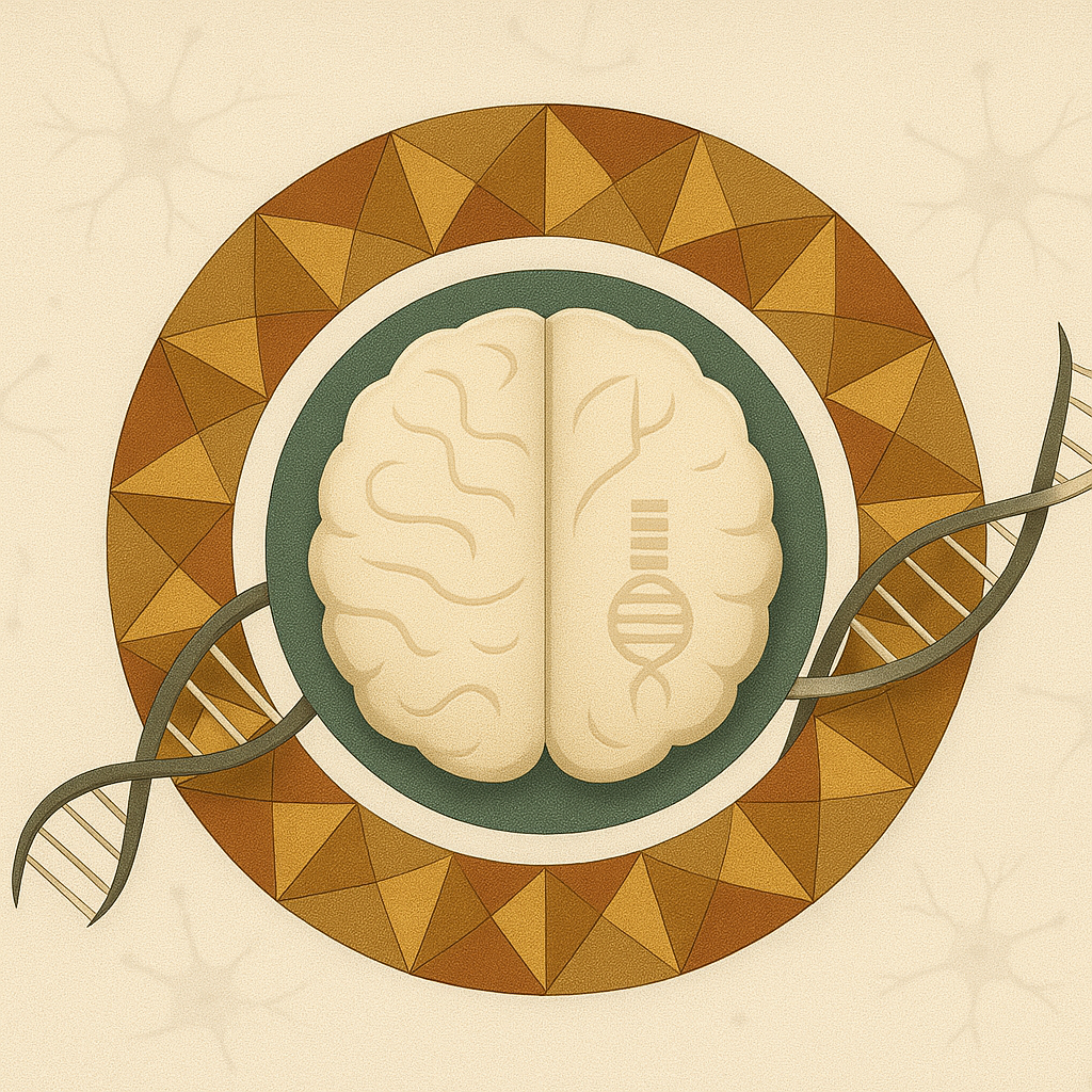
Amish lethal microcephaly, or MCPHA, is a severe genetic disorder that leads to an abnormally small head and brain. It is an autosomal recessive condition, meaning a child must inherit a copy of the flawed gene from both parents to be affected. Found almost exclusively within Old Order Amish communities in Pennsylvania, it occurs in approximately 1 out of every 500 births.
The condition is characterized by profound brain malformations that are present at birth and a prognosis that is unfortunately fatal, with death typically occurring within the first year of life. This article will explain the chain of events that causes this devastating disease, beginning with a single error in the genetic code and ending with catastrophic failure in brain development.
The Genetic Origin: A Flaw in the SLC25A19 Gene
The root cause of MCPHA is a mutation in a single gene known as SLC25A19. This gene contains the instructions for building a vital protein that acts as a transport vehicle. Its primary job is to carry thiamine pyrophosphate (TPP), the active form of vitamin B1, into the mitochondria—the microscopic powerhouses inside our cells.
In individuals with MCPHA, a specific and particularly destructive typo occurs in the SLC25A19 gene. This "missense mutation" causes the wrong protein building block to be used, which cripples the resulting transport protein. This single error results in a catastrophic failure to deliver TPP, setting off a deadly cascade of events within the cell.
The devastating nature of this specific genetic flaw is highlighted when compared to other mutations in the same gene. In non-Amish populations, different mutations in the SLC25A19 gene cause a related but less severe and treatable condition. This contrast underscores just how uniquely damaging the specific mutation responsible for MCPHA is.
The Cellular Crisis: How the Genetic Flaw Shuts Down the Brain's Power Supply
To understand how one genetic error leads to such a tragic outcome, we must look inside the cell's energy factories: the mitochondria. These organelles are responsible for generating the vast majority of the energy needed for survival and growth, a process especially critical for the rapidly developing brain. But how does the MCPHA mutation trigger a full-blown crisis at this cellular level?
An Energy Factory Grinds to a Halt
The delivery of TPP is essential because it acts as a crucial helper molecule for several key enzymes that drive the Krebs cycle, the central pathway of cellular energy production. Without enough TPP, these enzymes, including pyruvate dehydrogenase and alpha-ketoglutarate dehydrogenase, cannot function.
This breakdown effectively shuts down the main assembly line for producing ATP, the cell's universal energy currency. The developing brain has massive energy demands, and this sudden, severe energy deficit is a blow it cannot withstand. The power supply is cut off at the most critical time, halting the fundamental processes of brain growth.
A Buildup of Toxic Waste
When the primary energy pathway is blocked, the problems multiply. Metabolic compounds that should be processed begin to accumulate to toxic levels, creating a state of cellular emergency.
The failure of the alpha-ketoglutarate dehydrogenase enzyme directly causes its target molecule, alpha-ketoglutarate, to build up in the blood and urine. High concentrations of this substance are known to be toxic to the central nervous system, disrupting the delicate chemical environment that brain cells need to survive.
At the same time, cells desperate for energy switch to a less efficient backup system that produces lactic acid as a byproduct. This leads to lactic acidosis, a dangerous condition where the blood becomes too acidic, impairing organ function and signaling a body-wide energy crisis.
The Final Result: Irreversible Brain Damage
The systemic energy failure and toxic buildup during fetal development leave a permanent and devastating imprint on the central nervous system. This impact extends far beyond a small head size, manifesting as severe structural brain abnormalities and profound neurological dysfunction.
The developing fetal brain is uniquely vulnerable. The creation of billions of neurons and the formation of trillions of connections is an incredibly energy-intensive process. When the energy supply is cut during this critical period, brain development is abruptly halted. This results in the condition's most prominent feature: a severely undersized brain, or microcephaly.
Brain imaging studies reveal the extent of the damage. Key features include:
- A Smooth Brain Surface: The cerebral cortex often appears abnormally smooth, a condition known as lissencephaly. This occurs because neurons failed to migrate to their proper locations to form the brain's characteristic complex folds.
- Underdeveloped Structures: The cerebellum, which controls posture and coordination, is often underdeveloped. In many cases, the corpus callosum—the main bundle of nerve fibers connecting the brain's two hemispheres—fails to form at all.
- Severe Neurological Impairment: From birth, infants show signs of widespread brain damage. They typically have low muscle tone in their trunk, which contrasts with rigid, increased muscle tone in their limbs. They also experience frequent and disruptive involuntary muscle twitches, known as myoclonus.
This malformed and metabolically stressed brain environment leads to severe, difficult-to-control seizures. The combination of intractable seizures, profound developmental arrest, and the underlying energy crisis makes survival beyond infancy impossible, cementing the condition's lethal designation.
