

What is X-linked Ectodermal Dysplasia?
X-linked hypohidrotic ectodermal dysplasia (XLHED) is the most common type of ectodermal dysplasia, a group of genetic disorders affecting the development of the ectoderm—the outer layer of an embryo. This layer is responsible for forming many of the body's external structures. Consequently, XLHED primarily disrupts the normal development of hair, teeth, and sweat glands.
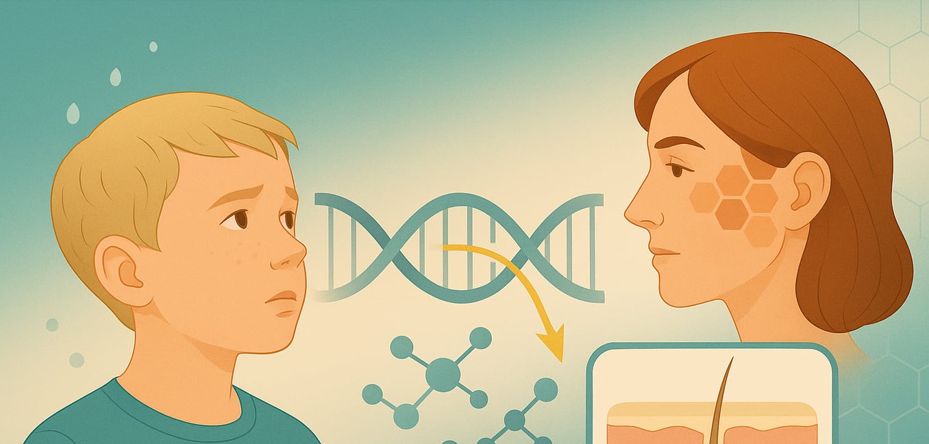
The condition is caused by a mutation in a gene located on the X chromosome, which is why it predominantly affects males. Signs of the disorder are often apparent from birth or early infancy and can range in severity. Understanding its specific signs, genetic roots, and unique inheritance pattern is key to diagnosis and effective management.
Signs and Symptoms
The signs of XLHED are often noticeable from birth and primarily impact structures derived from the embryonic ectoderm. While the severity can vary, affected males typically show a fuller spectrum of symptoms than female carriers.
The Core Triad: Hair, Teeth, and Sweat Glands
The condition is most recognized by its impact on three key areas. The most critical symptom is a severely reduced or absent ability to sweat (hypohidrosis or anhidrosis) due to underdeveloped sweat glands. Because sweating is the body's primary cooling mechanism, this inability can lead to dangerously high body temperatures (hyperthermia), which can be life-threatening, especially for infants. Hair is typically sparse, fine, brittle, and slow-growing (hypotrichosis), affecting the scalp, eyebrows, and eyelashes. Dental development is also profoundly affected, with many individuals having multiple missing teeth (hypodontia) or teeth that are small, conical, or peg-shaped.
Distinctive Facial and Skin Features
Individuals with XLHED often share a set of distinctive facial characteristics. These can include a prominent forehead (frontal bossing), a flattened or depressed bridge of the nose often described as a "saddle nose," and full, prominent lips. The skin is commonly thin, pale, and chronically dry, which can lead to conditions like eczema. A particularly notable feature is the skin around the eyes, which may appear thin, finely wrinkled, and darker in color (periorbital hyperpigmentation), a sign that can be present even in young children.
Respiratory and Glandular Effects
The disorder's effects extend to mucous membranes due to the defective development of various exocrine glands. This can cause chronic dryness of the eyes, mouth, and airways, leading to a heightened susceptibility to respiratory infections, as the glands that normally help clear pathogens are underdeveloped. Other related symptoms may include a chronic cough, a hoarse voice, and a foul-smelling discharge from the nose (ozena). While physical development is impacted, intellectual ability and overall growth are typically normal.
The Genetic Cause
The root of X-linked hypohidrotic ectodermal dysplasia lies in a mutation within a specific gene that orchestrates the development of an embryo's outer layer. This gene provides essential instructions for forming ectodermal structures.
The Role of the EDA Gene
As the name of the disorder suggests, the primary cause of XLHED is a mutation in the EDA gene, which is located on the X chromosome. This gene holds the instructions for making a crucial protein called ectodysplasin-A. During embryonic development, this protein acts as a key messenger, facilitating communication between two fundamental cell layers: the ectoderm and the mesoderm. This signaling is essential for the proper formation of sweat glands, hair follicles, and teeth. When a mutation occurs in the EDA gene, the resulting protein is either faulty or not produced at all. This disrupts the vital communication pathway and leads directly to the hallmark symptoms of the disorder.
Other Related Genetic Causes
While mutations in the EDA gene are responsible for the X-linked form, other, less common forms of hypohidrotic ectodermal dysplasia exist. These are caused by mutations in different genes, such as EDAR, EDARADD, and WNT10A. These forms follow different inheritance patterns (autosomal dominant or recessive) but result in very similar physical symptoms because they disrupt related biological pathways involved in ectodermal development.
How is it Inherited?
XLHED is passed down through families according to specific genetic rules that explain why males and females experience the condition differently. The inheritance pattern is dictated by the location of the mutated gene on the X chromosome.
X-linked Recessive Inheritance
The disorder follows an X-linked recessive pattern because the responsible EDA gene is on the X chromosome. Males have one X and one Y chromosome (XY). Therefore, inheriting a single copy of the mutated EDA gene on their lone X chromosome is sufficient for them to develop the full condition. A key characteristic of this pattern is that an affected father cannot pass the disorder to his sons, as he contributes his Y chromosome to them. However, he will pass the mutated gene to all of his daughters, who will then become carriers.
The Role of Female Carriers
Females have two X chromosomes (XX). If they inherit one mutated EDA gene, they are considered carriers. They typically experience milder and more variable symptoms than males due to a natural process called X-inactivation, where each cell randomly deactivates one of its two X chromosomes early in development. This results in a "mosaic" of cells—some using the healthy X chromosome and others using the X with the mutated gene. This explains why a carrier might have patches of skin that cannot sweat, a few missing teeth, or sparse hair. Approximately 70 percent of female carriers show some signs of the condition.
Other Inheritance Patterns
In less frequent cases where HED is caused by mutations in genes not on the sex chromosomes (like EDAR or WNT10A), the inheritance is autosomal. In an autosomal dominant pattern, only one copy of the mutated gene is needed to cause the disorder. In an autosomal recessive pattern, a person must inherit two copies—one from each parent—to be affected. These patterns affect males and females equally.
Onset and Diagnosis
Diagnosing XLHED often begins when an infant or young child shows the first signs of the condition. The process involves clinical observation, a review of family history, and confirmation with genetic testing.
Initial Clinical Evaluation
The diagnosis is frequently suspected by a pediatrician based on a physical examination. A critical early indicator can be recurrent, unexplained high fevers in an infant, which points to a reduced ability to sweat. A doctor will also look for other characteristic features, including very fine or sparse hair on the scalp and eyebrows, and distinctive facial traits like a prominent forehead and a flattened nasal bridge.
The Importance of Dental Assessment
A thorough dental evaluation is a cornerstone of the diagnostic process. A pediatric dentist can identify tell-tale abnormalities, such as multiple missing teeth or existing teeth that are small and conical. A panoramic X-ray is often used to get a clearer picture, allowing the dentist to see beneath the gums and confirm the absence of developing permanent tooth buds. This finding is a strong indicator of ectodermal dysplasia and is crucial for planning future dental care.
Confirmation with Genetic Testing
For a definitive diagnosis, genetic testing provides the final confirmation. A simple blood test is used to analyze DNA and identify a mutation in the EDA gene. This molecular testing is invaluable for confirming the diagnosis in an affected male, identifying female carriers within the family, and distinguishing XLHED from other forms of HED caused by different genes. This ensures the correct diagnosis and allows for appropriate genetic counseling.
