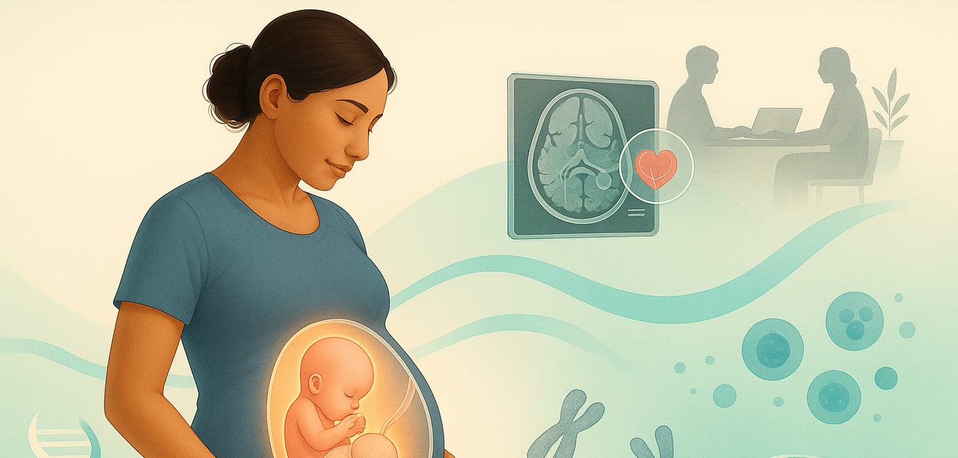


What is Omphalocele? A Brief Overview
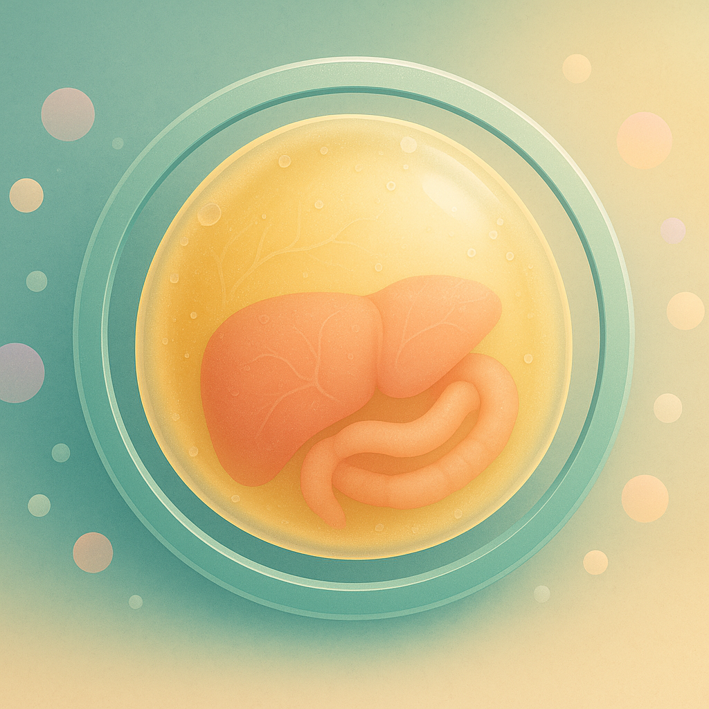
An omphalocele is a type of abdominal wall birth defect where a baby's organs, such as the intestines and liver, protrude through an opening at the base of the umbilical cord. These organs are not exposed to the amniotic fluid; they are contained within a thin, protective sac.
This condition occurs very early in pregnancy. Between the sixth and tenth weeks of fetal development, it is normal for the intestines to temporarily bulge into the umbilical cord as they grow rapidly. By the eleventh week, they typically return to the abdominal cavity. An omphalocele results when this final step does not happen, leaving some organs outside the body at birth.
The Diagnostic Pathway: From Initial Scan to Detailed Assessment
The diagnosis of omphalocele is a step-by-step process that begins with a routine scan and progresses to more specialized evaluations. This pathway is designed to confirm the diagnosis, determine its severity, and check for any associated health conditions.
Initial Detection with Prenatal Ultrasound
An omphalocele is most often first identified during a routine second-trimester anatomy scan, usually performed around 20 weeks of pregnancy. While a bulge of intestines into the umbilical cord is normal before 11 weeks, the persistence of organs outside the abdomen after this point suggests an omphalocele.
During this high-resolution scan, a sonographer will look for specific signs:
- A sac protruding from the belly button where the umbilical cord inserts.
- The presence of a thin, protective membrane covering the organs.
- Identification of which organs, like the liver or bowel, are inside the sac.
This detailed ultrasound also serves as a comprehensive screening of the baby’s entire body to check for other potential abnormalities, providing the first complete picture of the baby’s health.
Advanced Imaging: Fetal MRI
Following an ultrasound, a fetal MRI may be recommended to provide an even more detailed view of your baby’s anatomy. This imaging technique uses a powerful magnetic field to create clear, cross-sectional images without using radiation.
An MRI is particularly useful for measuring the baby's lungs. By comparing your baby’s lung size to the expected size for their gestational age, doctors can predict whether the baby might need breathing support after birth. This advanced knowledge helps the medical team prepare the right equipment and care plan for delivery.
Assessing the Heart: The Fetal Echocardiogram
Because heart defects are frequently associated with omphalocele, a fetal echocardiogram is a critical part of the diagnostic process. This is a specialized, in-depth ultrasound that focuses exclusively on the baby’s heart.
Performed by a pediatric cardiologist, this scan provides a moving picture of the heart's chambers, valves, and major blood vessels. It assesses both the heart's structure and its function, ensuring blood is pumping correctly. Identifying a heart condition before birth is crucial, as it allows the medical team to create a precise, coordinated plan and ensure the right specialists are ready to provide immediate care after delivery.
Investigating Associated Genetic Conditions
Discovering an omphalocele often leads to an evaluation of your baby’s genetic makeup, as the condition can be linked to certain chromosomal abnormalities or genetic syndromes. Understanding these potential links helps create a comprehensive care plan that addresses all aspects of your baby’s health.
The Role of Genetic Counseling
A genetic counselor is a key member of your care team who helps you navigate the complexities of genetic testing. This specialist will discuss your family and medical history, explain the available tests, and detail what those tests can and cannot reveal. They provide expert guidance and support, ensuring you can make informed decisions about your baby’s care.
Diagnostic Testing with Amniocentesis
To get definitive answers about your baby’s genetic health, your doctor may recommend an amniocentesis. This diagnostic test, typically performed between 15 and 20 weeks of pregnancy, involves collecting a small sample of the amniotic fluid that surrounds the baby.
This fluid contains fetal cells with your baby’s unique genetic information. Analyzing these cells allows for a detailed examination of the chromosomes to check for conditions such as trisomy 13, 18, and 21, as well as other genetic syndromes. The results are a vital piece of the puzzle, allowing the medical team to anticipate other health needs and assemble the right specialists before your baby is born.
