


How Healthcare Teams Assess the Severity of an Omphalocele
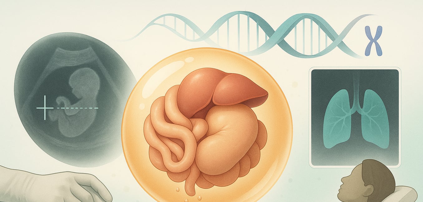
Omphalocele is a birth defect of the abdominal wall where a baby’s internal organs protrude through the base of the umbilical cord. This occurs early in pregnancy when the abdominal wall fails to close completely. Unlike a similar condition, gastroschisis, the organs in an omphalocele are contained within a thin, protective membranous sac.
While the diagnosis itself is straightforward, the condition’s severity varies widely. A thorough and multi-faceted assessment is critical for predicting a baby's prognosis and creating a comprehensive care plan. This evaluation process moves beyond simply identifying the defect to carefully analyzing its physical traits, using advanced imaging to understand its impact on other organs, and investigating for associated genetic conditions.
Component 1: Evaluating the Physical Defect
The initial assessment focuses on the physical characteristics of the omphalocele itself. The size of the opening, the specific organs involved, and the integrity of the protective sac are all critical factors that help determine the complexity of the case.
Size and Contents
Omphaloceles are generally categorized as either small or "giant." A small defect may only contain a portion of the intestines and can often be repaired with a single surgery after birth.
A giant omphalocele, typically defined as a defect larger than five centimeters, presents a far greater challenge. These larger defects often contain not only the intestines but also the stomach, spleen, and a significant portion of the liver. This leads to a major issue known as visceral-abdominal disproportion. In simple terms, this means the baby’s abdominal cavity is underdeveloped and too small to hold all the herniated organs at once, requiring a more complex, staged surgical repair.
The Critical Role of the Liver
The single most important factor in assessing an omphalocele’s severity is whether the liver is inside or outside the abdomen. When the liver is part of the herniated organs, it significantly increases the risks and complexity of management.
A cesarean section is often recommended to prevent the large, fragile liver from being damaged during delivery, which could cause life-threatening bleeding. Surgically, returning a large, solid organ like the liver to a small abdominal cavity is much more difficult than returning loops of bowel, often necessitating a delayed, multi-stage repair process.
The Protective Sac
The thin, translucent sac covering the organs is a key feature of an omphalocele. Made of the peritoneum and amnion, this membrane provides a vital barrier, protecting the organs from infection and injury.
In rare cases, this sac can rupture before or during delivery. A ruptured omphalocele requires immediate and urgent medical care to prevent infection, dehydration, and heat loss. The exposed organs must be immediately wrapped in sterile dressings, and the baby often requires more urgent surgical intervention.
Component 2: Comprehensive Imaging for a Deeper Look
To get a complete picture of the baby’s health, specialists rely on a combination of imaging techniques. These tools provide essential information about the omphalocele, associated organ function, and potential complications that will guide care both before and after birth.
Foundational Imaging: Ultrasound and Echocardiogram
A high-resolution fetal ultrasound is the cornerstone of prenatal diagnosis. It is used to confirm the omphalocele, measure its size, and identify which organs—especially the liver—are inside the sac. Ultrasound is also used to screen for other physical abnormalities that may be present.
Due to the strong link between omphalocele and congenital heart defects, a fetal echocardiogram is a standard part of the evaluation. This specialized ultrasound provides a detailed view of the baby’s heart structure and function. Identifying a heart condition before birth allows a full cardiac team to be prepared, ensuring the baby receives immediate, life-sustaining care at delivery.
Advanced Fetal MRI for Lung Assessment
A serious complication, particularly with giant omphaloceles, is pulmonary hypoplasia, which simply means underdeveloped lungs. When organs are displaced into the umbilical cord, the abdominal cavity is smaller, which can restrict chest growth and prevent the lungs from developing fully. The severity of pulmonary hypoplasia is a primary predictor of a baby’s survival and respiratory health.
To assess this risk, many specialized centers use fetal magnetic resonance imaging (MRI). This advanced imaging provides precise measurements of lung size and helps predict how much breathing support a baby may need after birth. Key metrics include:
- Total Lung Volume (TLV): This is a direct measurement of the baby’s lung size. Research has shown that a very small TLV is linked to a higher need for breathing support, such as intubation, immediately after birth.
- Observed-to-Expected (O/E) Lung Volume: This ratio compares the baby’s measured lung volume to the average volume expected for a healthy fetus of the same gestational age. A low O/E TLV ratio is a powerful predictor of severe pulmonary hypoplasia, longer hospital stays, and overall survival rates.
This detailed information helps the medical team counsel families and plan the safest delivery and surgical strategy. For instance, if lungs are severely underdeveloped, surgeons may opt for a delayed repair to allow the baby’s body and lungs time to grow.
Component 3: Investigating Associated Health Conditions
An omphalocele is often a visible sign of other, less obvious health issues. A thorough investigation for associated anomalies and underlying genetic factors is essential, as these co-occurring conditions can be more significant for the baby's long-term health than the omphalocele itself.
Chromosomal Abnormalities
There is a strong association between omphalocele and trisomies, which are conditions caused by an extra chromosome. The most common are Trisomy 18 (Edwards syndrome) and Trisomy 13 (Patau syndrome). Both are associated with severe, multi-system health problems and have a very poor prognosis. When an omphalocele is linked to one of these conditions, the focus of care and family counseling shifts to address the life-limiting nature of the underlying genetic disorder.
Beckwith-Wiedemann Syndrome (BWS)
BWS is the most common genetic syndrome associated with omphalocele. It is an overgrowth disorder with several key characteristics, including:
- A large body size at birth (macrosomia).
- An enlarged tongue (macroglossia).
- Low blood sugar in the newborn period (neonatal hypoglycemia).
A critical aspect of managing BWS is the increased lifetime risk of developing certain childhood cancers, particularly Wilms tumor (a kidney cancer) and hepatoblastoma (a liver cancer). Because of this, children with BWS require a long-term screening protocol with regular blood tests and abdominal ultrasounds for many years.
Other Structural Defects
Even without a specific syndrome, babies with an omphalocele may have other structural malformations. As mentioned, heart defects are particularly common. Other potential issues can involve the central nervous system, spine, and kidneys. Identifying these additional anomalies is vital for creating a truly comprehensive treatment plan that addresses all of the baby's health needs.
